

What if the treatment for your cancer was as unique as you are?
That’s the promise of a new technology developed at the Technion by doctoral students Gil Shamai and Ron Slossberg and Professor Ron Kimmel of the Technion Faculty of Computer Science, in collaboration with Dr. Yoav Binenbaum of Ichilov Hospital and Professor Ziv Gil of Rambam Medical Center.
The deep learning–based method for mapping critical receptors on cancer cells is expected to lead to better, more personalized cancer treatment.
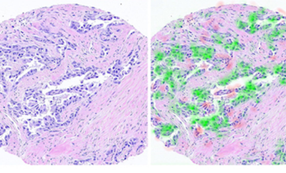
The technology extracts molecular information from digital biopsy images that underwent hematoxylin and eosin (H&E) staining. H&E staining allows the pathologist to identify the type of cancer and its severity in the tissue under the microscope.
But staining can’t tell us everything. Understanding the molecular profile of the tumor, its biological pathways, the genetic code of the cancer cells, and the common receptors on the cell membrane are essential in developing the right treatment for a patient.
Using Deep Learning in Cancer Care
So the Technion researchers found a better way, extracting molecular information from the cell shape and the environment (known as the morphology of the tissue) as reflected in the H&E scans.
With so many variables in a tumor’s shape, many pathologists assumed it was impossible to determine a tumor’s features based on shape. But by using deep learning and other artificial technologies, the Technion researchers are able to characterize the cancer with a complex analysis of its morphology…by simply looking at the tissue as it appears on a standard H&E scan.
It’s not easy to build a deep-learning system. The computer needs a huge amount of information to analyze and identify patterns. The Technion researchers accelerated the innovation process by writing a software code to scan network sources and automatically download thousands of biopsy samples and the relevant medical information approved for research.
Prof. Kimmel and the team examined more than 20,000 scans from 5,356 breast cancer patients. Using the new technology, they were able to map estrogen and progesterone receptors, among other molecular biomarkers, from the scans alone and based on cell morphology.
A Promising New Tool
While the team’s research focused on breast cancer, it is clear this technology could be used for all cancers: first as a decision-making tool to aid doctors, and later as a clinical tool for diagnostic purposes.