

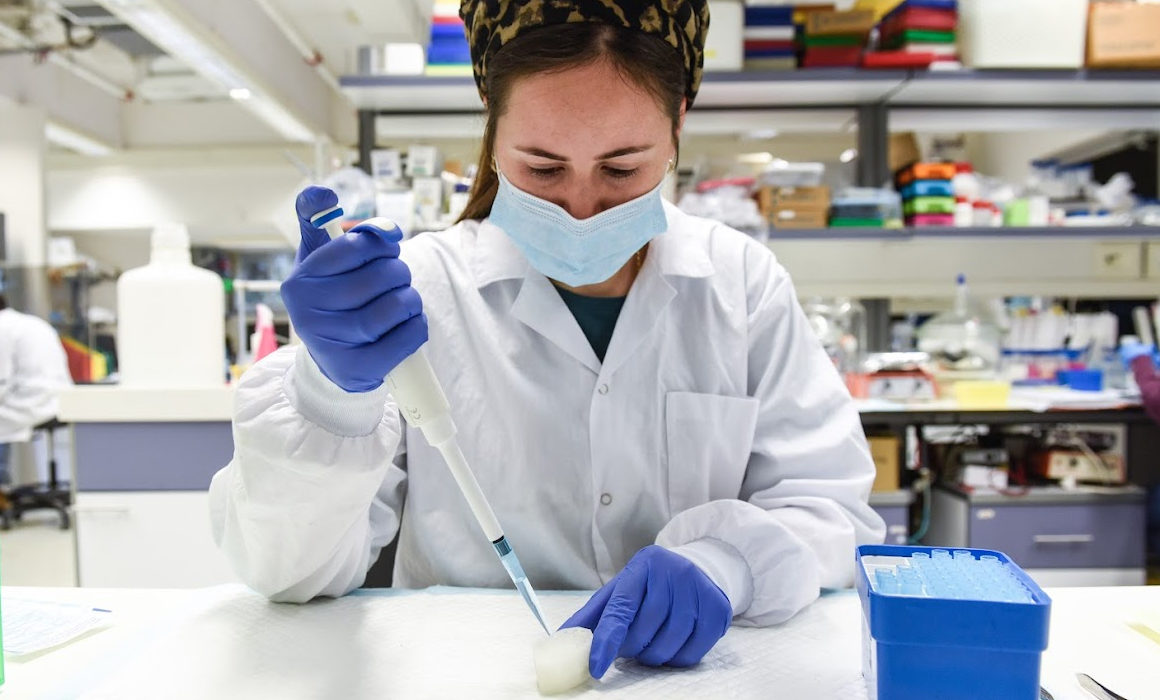
Pictured Above / Dr. Shira Landau
Researchers at the Technion – Israel Institute of Technology and Sheba Medical Center have developed a technology that can efficiently make custom-made functional aesthetic implants for patients with microtia, or congenitally deformed ears.
Microtia is a birth defect that occurs when the external ear fails to develop normally, and as a result, is small and improperly formed. Microtia occurs in 0.1 to 0.3 percent of births. Occasionally, besides the aesthetic issue, microtia also involves hearing loss.
Since the “bones” of the outer ear — the auricle — are in fact flexible cartilage and not bone tissue, the customary technique for microtia reconstruction is to use costal cartilage harvested from the patient’s chest. This process is extremely painful and risks additional complications. Moreover, constructing an ear that is identical to the other one depends on both the surgeon’s creativity and high-level surgical skills.
The breakthrough is the result of collaboration between Professor Shulamit Levenberg of the Faculty of Biomedical Engineering at the Technion and Dr. Shay Izhak Duvdevani, a senior physician in the Otorhinolaryngology Head and Neck Surgery Department and Head of the Tissue Engineering Lab at Sheba Medical Center. The researchers applied new technologies for tissue engineering, developed in Prof. Levenberg’s lab under the leadership of Dr. Shira Landau, to fabricate a biodegradable auricle scaffold that formed stable, custom-made neocartilage implants.
The unique scaffold is 3D-printed and based on a CT scan. It is biodegradable and forms chondrocytes — the cells responsible for cartilage formation — and mesenchymal stem cells. The scaffold has pores of varying sizes, allowing for cell attachment to form stable cartilage.
According to the researchers, engineering an auricle from the patient’s own cells will reduce the suffering and risk caused to children as a result of harvesting their costal cartilage. Furthermore, it will allow the surgery to be performed on children as young as six years old, rather than the currently accepted practice of waiting until they are ten. Performing the surgery at a younger age is likely to mitigate the psychological effects of microtia on children.
According to Prof. Levenberg, “One of the challenges in the study was to find a suitable 3D printing method, since fabricating an ear necessitates the use of biodegradable materials that break down in the body without harming it but have an extremely accurate external structure and small pores. We demonstrated all of this in the present research, and estimate that it will be possible to tailor our technology to other applications, such as nasal reconstruction and fabrication of various orthopedic implants.”